Your Partner In Digital Success


YOUR PARTNER IN DIGITAL SUCCESS,
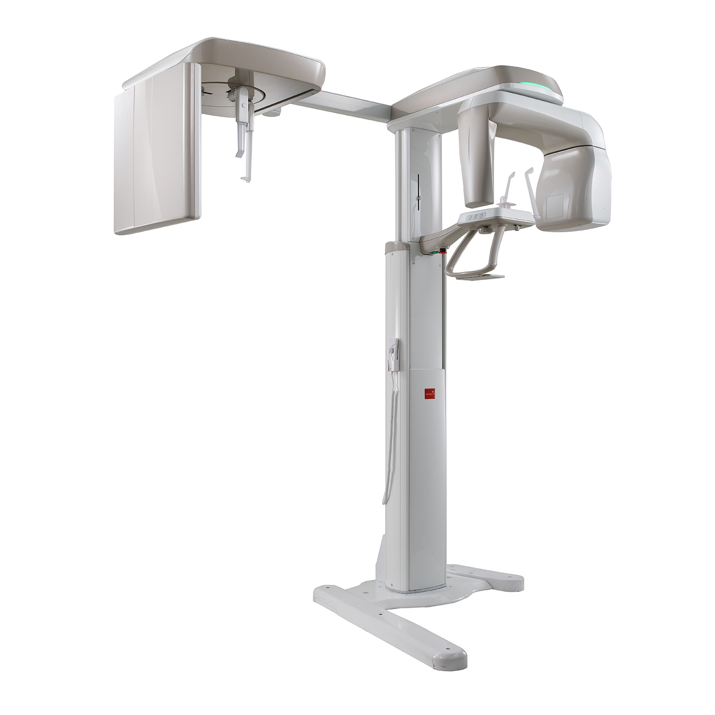
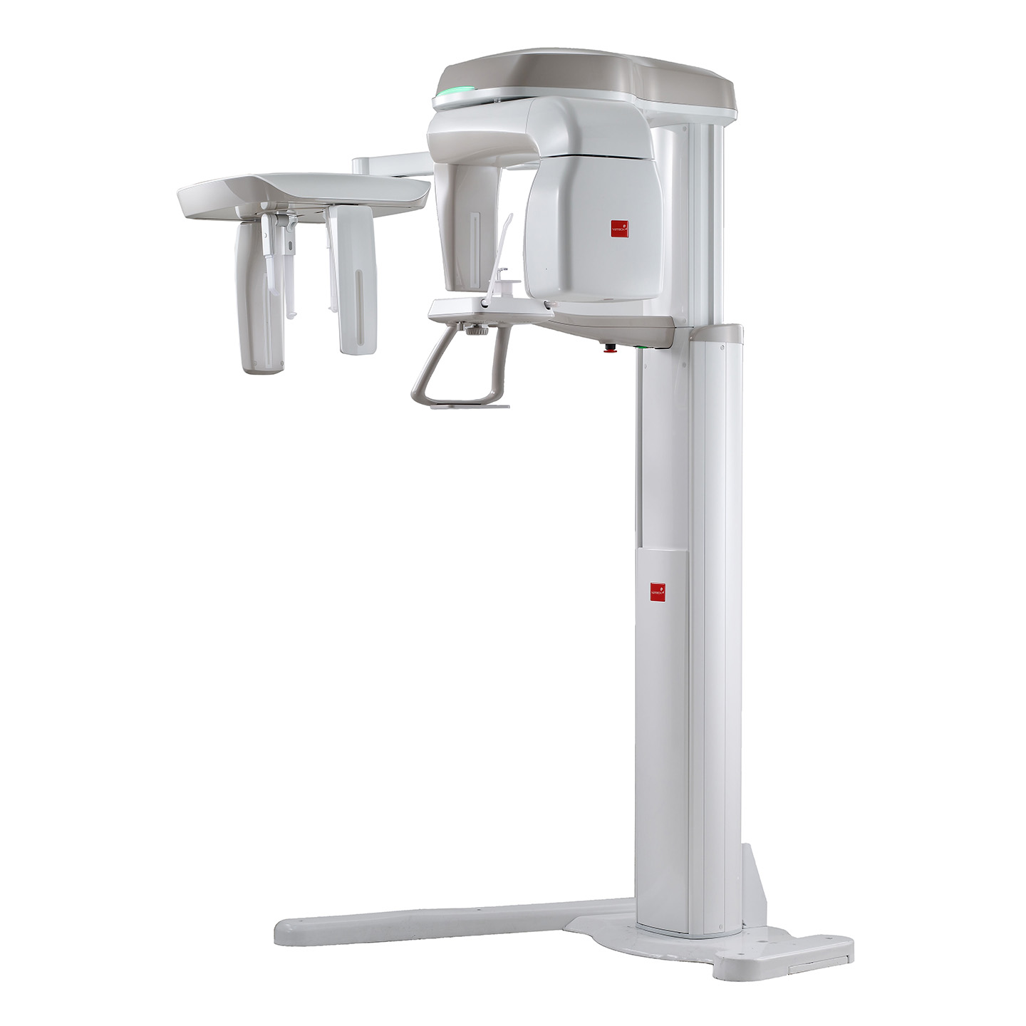
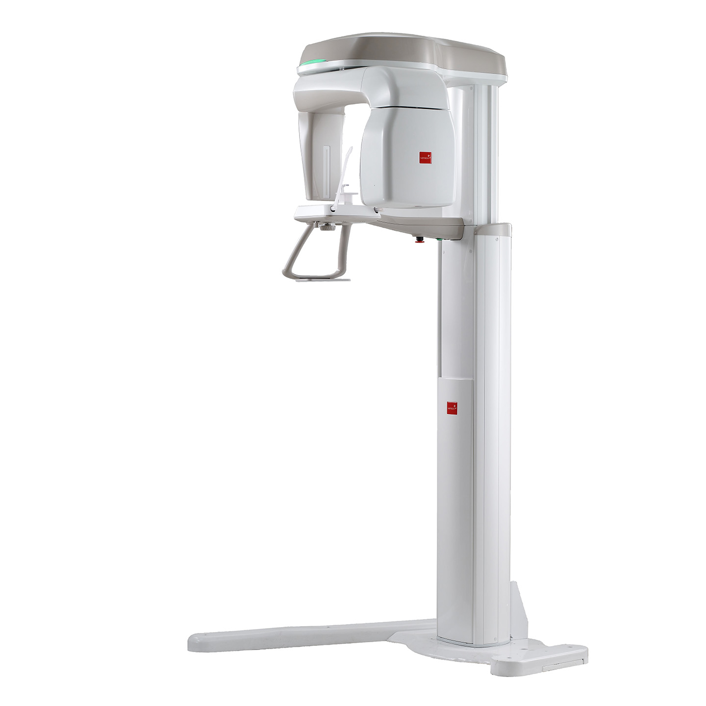
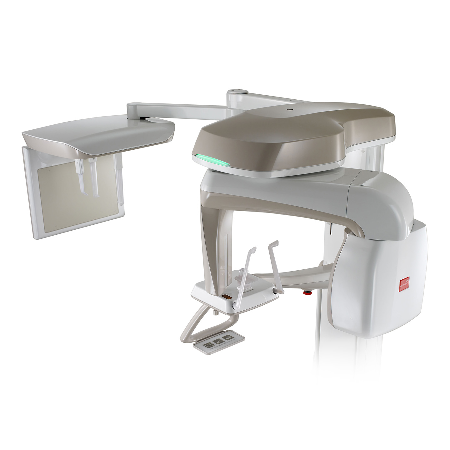
PaX-i
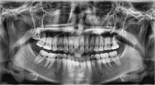
| Superior Image Quality |
|
|---|---|
| Two Dedicated Sensors |
|
| Modern and Compact Design |
|
THE ADVANCED IMAGING SOLUTION FOR ACCURATE DENTAL DIAGNOSIS
The PaX-i provides the most precise and high quality panoramic images by combining image
processing and accumulated experience in dental imaging from Vatech.
This will increase your diagnostic accuracy for improved treatment planning and
patient satisfaction.
MAKE YOUR DIAGNOSIS EASY AND EFFICIENT WITH VARIOUS CAPTURE MODES
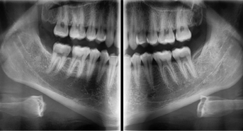
Bitewing Mode
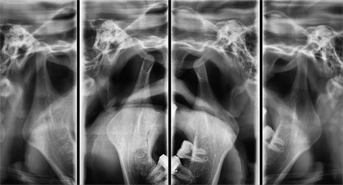
TMJ Mode
| SELECTION | ARCH | EXAMINATION MODE |
|---|---|---|
| PANO EXAMINATION | Narrow / Normal Wide / Child | Standard / Right / Front / Left |
| Orthogonal | Orthogonal Standard / Right / Front / Left Bitewing Standard / Right / Front / Left | |
| SPECIAL EXAMINATION | Normal | TMJ LAT Open / Close TMJ PA Open / Close Sinus LAT / PA |
CEPHALOMETRIC (SCAN CEPH)
The PaX-i provides optimal images exclusively designed for orthodontics.
There are two image sizes available, Lateral and Full Lateral, allowing you to choose your image size based on your diagnostic needs.
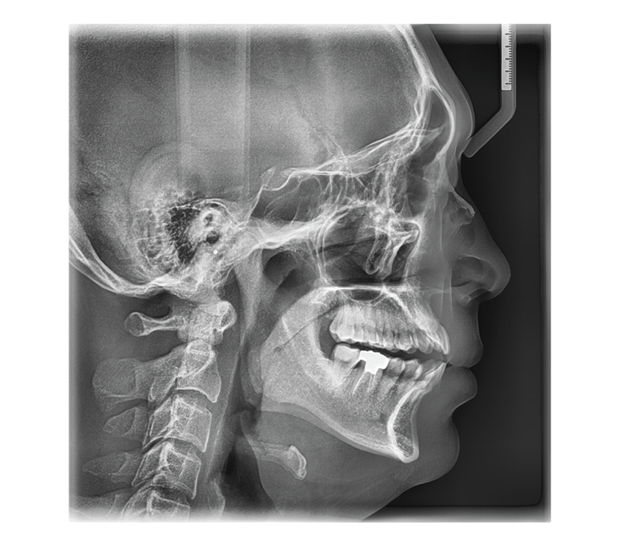
LATERAL
Provides specialized high quality images to suit orthodontics and maxillofacial surgeries.
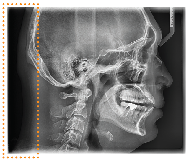
FULL LATERAL
A full lateral image size is 30% wider and shows the occipital area of the patient, which enables comprehensive diagnosis.
CEPHALOMETRIC (ONE SHOT TYPE)
Superior image quality will be delivered using the a-Si TFT sensors. Three different ceph image sizes reduce unnecessary x-ray dosage and scan the ideal area of cranial anatomy for your diagnosis and treatment planning.
LATERAL

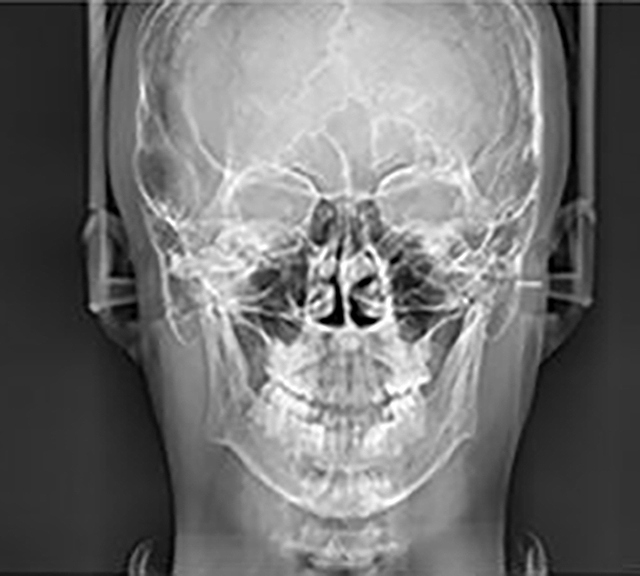
PA
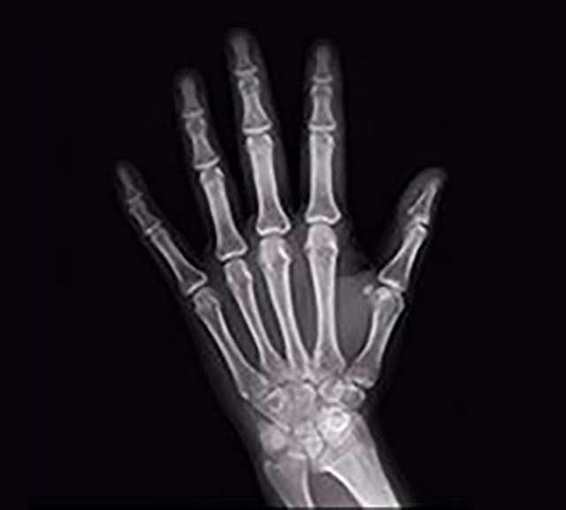
Carpus
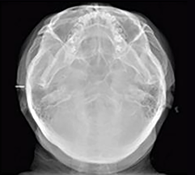
SMV (Submentovertex)
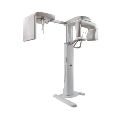
PRODUCT CONFIGURATION
| PANO | CEPH | ||
|---|---|---|---|
| SCAN | ONE SHOT | ||
| Pax-i | ● | - | - |
| Pax-i SC | ● | ● | - |
| Pax-i OP | ● | - | ● |
SPECIFICATIONS (PaX-i : PCH-2500)
| Function | Pano + Ceph |
|---|---|
| Scan Time |
Pano : HD 13.5 sec / Normal 10.1 sec |
| Focal Spot | 0.5 mm |
| Tube Voltage / Current | Pano : 50~90 Kvp / 4-10 mA |
| Ceph FOV Size |
SC |
| Gray Scale | 14 bit |
| Patient Positioning | Standing / Wheel-chair accessible |
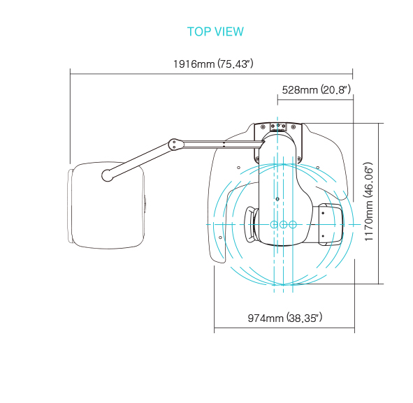
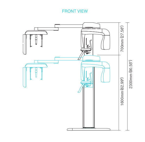
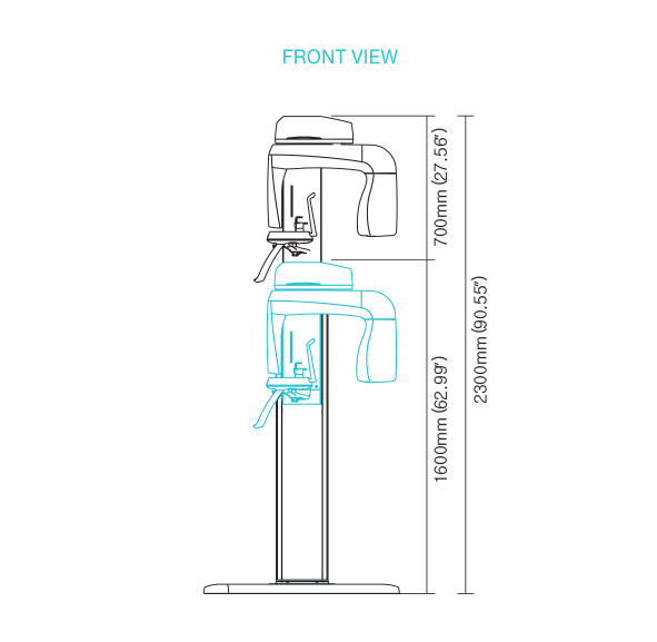
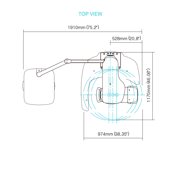
DIMENSIONS [Unit: mm]
Pano
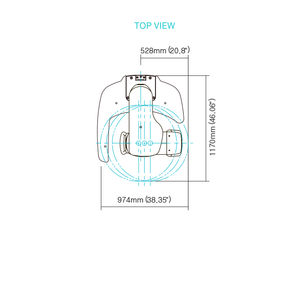
Pano/Scan Ceph
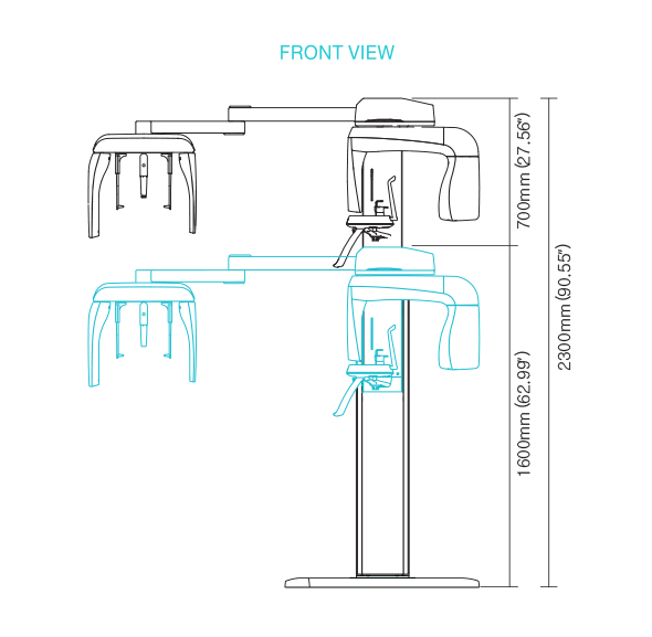
Pano/OneShotCeph